


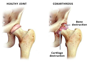
Coxarthrosis - degenerative disease that leads to destruction of the hip joint and is a chronic. More common in older age groups. More common in women than in men.
Begins gradually and progresses slowly. Can affect one joint or both. Is the most common is osteoarthritis.
Why is the disease?
Osteoarthritis in some patients is involved with the natural aging process is the degeneration of the tissues of the hip joint. Its appearance is affected by several factors:
- reduces tissue nutrition;
- congenital abnormality of the hip joint, in particular, dysplasia;
- trauma to the pelvis;
- post-infectious hip;
- aseptic necrosis of the head of the hip joint;
- Perthes disease (osteochondropathy).
Unfortunately, to find out the cause of the disease is not always possible and the pathology of the hip joint is called idiopathic coxarthrosis - it is so, the cause of which is not established. This is an incentive to continue the study of the problem. Scientific works in this field, and the doctors came to the conclusion that a higher risk of osteoporosis observed in the following patients:
- Hereditary predisposition to pathology. Patients whose parents suffer from diseases of cartilage and bone, in most cases, you also have a such problems;
- Overweight. A significant weight load on the joints, which are regularly exposed mechanical work;
- Metabolic disorders, diabetes. This leads to poor oxygen and nutrients to the joint tissue, causing them to lose their properties.
Knowing the main risk factors of the disease, and to design preventive measures to prevent it.
How to identify the pathology of the hip joint?
Symptoms of osteoarthritis depends on the anatomical features of the musculoskeletal system, causes of pathology and the stage of the process. Consider the main clinical symptoms:
- joint pain;
- irradiation of pain in the knee, thigh to the groin;
- the stiffness of the movement;
- limited mobility;
- violations to walk, limp;
- calculate the mass of thigh muscles;
- the shortening of the limb.
The clinical picture corresponds to the internal changes in the tissues of the joint. The symptoms increase gradually and in the early stages, the patient does not pay them enough attention. This is dangerous, because at the beginning of the treatment process will bring a greater impact.
Clinical and radiological degree of osteoarthritis
Listed below are the symptoms of each degree.
- 1 degree. The patient experiences intermittent pain and discomfort. Discomfort bother you after exercise, a long position in a static pose. Pain in the localized area of the joint and passing it after a rest. In this stage of the process is not impaired to walk and no shortening of the leg. The changes seen on radiographs - joint space narrowing, is the osteophytes (bony growths).
- Of 2 degrees. Increases the intensity of the pain, it can occur at rest and radiates to the nearby areas of the body. Seems to meet, after the man had walked or wave. Limited range of motion of the joint. Simultaneously, the change in x-ray images: the displaced head of the femur, osteophytes grow on the inner and outer edges of the acetabulum.
- Step 3. The pain becomes permanent, will come during the day and at night. Much worse to walk becomes a permanent lameness. Drastically reduced motor skills, atrophy of the leg muscles. changing the muscle tissue causes the legs slightly pulled up and becomes shorter. This leads to the deformation of the posture and curvature of the body. X-rays in this stage of the process: total narrowing of the gap between the surface of the joint deformity of the femoral head, significant increase in osteophytes.
The diagnostic program, when the disease
The main method of diagnosis - x-ray. It can be used to determine the presence of the disease and its stage. Radiography to analyze the structure of a common subject, narrowing of the joint space, osteophytes, fracture of the head of the hip bone.
If there is a need to study the condition of soft tissue MRI is performed. It allows you to study in detail the condition of the cartilage areas of the joint and the muscles of the hip area.
Modern methods and ways of treatment of coxarthrosis of the hip joint
Osteoarthritis treatment can be conservative and surgical. In the treatment of osteoarthritis aims at the following objectives:
- to reduce the pain symptoms;
- the restoration of the operation of the engine;
- rehabilitation and rehabilitation;
- prevention of complications;
- improve the quality of life of the patient.
Initiation of treatment is modification of risk factors. You can do this, the doctor recommends the following actions:
- normalization of weight;
- avoiding harmful habits;
- nutrition;
- normalization and physical activity;
- balanced drinking system;
- healthy sleep.
Conservative treatment are: drug and non-drug. Drug therapy includes non-steroidal anti-inflammatory drugs, analgesics, chondroprotectors. They reduce inflammation in the tissue, remove swelling and soreness, restore range of motion and improve the condition of cartilage.
Non-drug treatment includes, among other things, massage the affected area. It stimulates the muscles that oppose their degeneration and the prevention of the shortening of the limb. Perfect and professional massage stimulates the blood circulation in the area of the joint, and this, in turn, leads to the normalization of metabolism in tissues. Please note that massage is not always useful when coxarthrosis - it is only made between exacerbations and at certain stages of the process. Configure it to your doctor may recommend massage techniques, the diversity of treatments and the duration of the course.
Obligatory condition of treatment is physical therapy. Is the prevention of contractures and progression of the disease. Exercises should be performed daily, only then will they have an impact. The exercises are selected individually and is prescribed by a doctor. Exercises to improve general health, reduce emotional disorders, to strengthen the forces of the body.
Physiotherapy is another method, which applies to coxarthrosis. It can be mud, medical baths and showers, magneto therapy. Used electro - and phonophoresis of medicinal substances.
If these methods of treatment brought no effect or has been applied to late - surgical treatment is necessary.
Surgical intervention with coxarthrosis
Surgical treatment is used when ineffectiveness of conservative method. This is especially true for the late diagnosis. Modern operational techniques and high-quality devices work allow to restore the structure and function of the joint, to restore people to the range of motion and normal quality of life. The most effective method of surgical treatment is joint replacement surgery.
Indications for surgery are:
- coxarthrosis 2-3 degrees;
- the lack of effect of treatment;
- total restriction of movement, walking.
The subjects, who are not allowed to perform the operation of:
- heart failure, kidney, the heart, the liver;
- mental illness;
- in the acute phase of the inflammatory process in the body.
For this purpose, the preoperative diagnosis. However, if it is possible to adjust the patient's preparation for surgery and after the intervention.
The action involves removing the diseased tissue and the prosthesis. There are different models of implants. Different methods of fastening the bone – cement and cement-free material, in which the prosthesis. All the features of the prosthesis and the intricacies of the surgical procedure can get information about consultation with the attending physician.
The recovery period after the surgical treatment
The first day after surgery, rehabilitation is carried out under medical supervision. First, he is to carry out passive movements, as the load is increased gradually. Walk the first time is allowed only with crutches, getting on the seat and squat.
Of course, for the first time after surgery, there are limitations to the load. Do not be afraid - because without action, these limits should be preserved to the end. A decrease in physical activity after surgical treatment, it is necessary to strengthen the position of the prosthesis, restoration of the integrity of the bones, healing wounds. 2 months should be left to the sports activities exercise, long walk and exercise. After complete recovery, the person returns to normal life, to do sports and outdoor activities.
The service life of the prosthesis: some companies tell you the survival rate of 90% for observation times up to 15 years.